
 |
 |
 |
 |
|||||
| Healthy and Ethically-Orientated Nutrition |
MOLECULAR MORPHOLOGY
 |
 |
 |
|---|
From 1986 to 2005, Univ.-Prof. Dr. Gerhard W. Hacker worked on the development and modification of new methods of analytical microscopy. These include various techniques of Immuncytochemistry or immunohistochemistry (in particular: immunofluorescence, enzyme-based permanent procedures such as immunperoxidase, peroxidase anti-peroxidase (PAP), streptavidin-biotin peroxidase and NTB-BCIP-streptavidin-biotin methods, Immunogold-Silver Staining) and in situ molecular biology procedures (in situ hybridization, in situ 3SR, and in situ PCR). With the development of Immunogold-Silver Staining (IGSS) methods, the introduction of the silver acetate autometallography to gain improved silver development, Nanogold-silver staining, gold-silver in situ PCR and the super-sensitivity Nanogold hybridization, Prof. Hacker and his team were able to introduce methodologies which for the first time allowed smallest quantities of specific proteins and peptides, and also the first time specific DNA gene and RNA sequences in microscopy with molecular sensitivity. A new field of research was born: Molecular Morphology, also leading to new and MedLine-cited scientific journals, the Journal of Molecular Morphology, later on changed to Applied Immunohistochemistry and Molecular Morphology. Application of these methods in cancer research, endocrinology and immunohistopathology led to some 150 scientific original papers, review articles and book chapters of Prof. Hacker on these subjects and the organization of renommated international Congresses, also including Atlantic City and Orlando (U.S.A.), Montreal (Canada), and Salzburg.
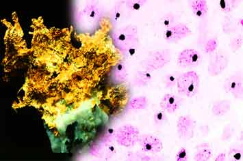
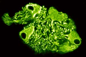
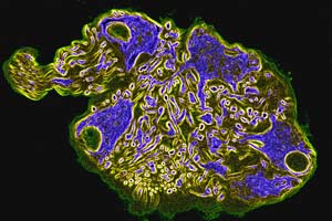
The above images show (at the far left) a gold nugget - a form of the basic natural mineral used to produce smallest particles of gold (Nanogold, with only 5 nanometers in diameter), which are manufactured at Nanoprobes, Inc., USA. In microscopy, such particles are used as Nanogold covalently linked to certain macromolecules, such as the Nanogold-streptavidin reagent. Using Nanogold, it is possible for example, to detect individual molecules of specific DNA sequences of human papillomavirus (HPV) 16/18 in an advanced squamous cell carcinoma of the cervix. In the photo on the left, the virus DNA sites can be seen as black spots distinctly standing out against the cancer cell nuclei counterstained in pink. Each of these black dots represents exactly one copy of a HPV-16/18 virus particle. The two pictures in the center and at the right show immunofluorescence preparations representing a small nerve cell collection with its corresponding cell processes, a ganglion in a urinary bladder of man – the center photo shows the usual green FITC fluorescence appearance, and the photo on the right is the result of computerized false-color staining allowing better visibility of nerve cell bodies.